Robotic Hernia Surgery: A New Era of Precision –
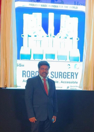
By Dr. Kumar- Billroth Hospitals
Hernia surgery has evolved dramatically over the last decade. From traditional open repairs to laparoscopic techniques, and now the most advanced form — Robotic Hernia Surgery. As a leading Hernia Specialist in Chennai, Dr. Kumar at Billroth Hospitals is among the few surgeons trained in new-generation robotic platforms, offering patients safer surgery, faster recovery and superior outcomes.
About Dr Kumar
Dr Kumar at Billroth Hospitals is an Advanced Laparoscopic and Robotic Hernia Surgeon with over 29+ years in the field of hernia surgery of Experience and Expertise of more than 10,000 Hernia Surgeries behind him. He Specializes in Laparoscopic and Robotic Hernia Surgeries, Complex and Recurrent Hernia Surgeries and Abdominal Wall
Reconstruction
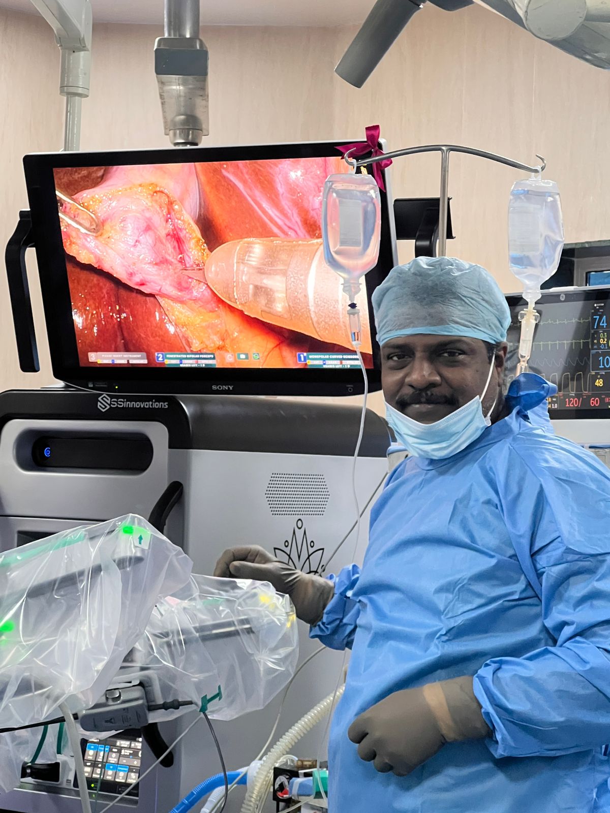
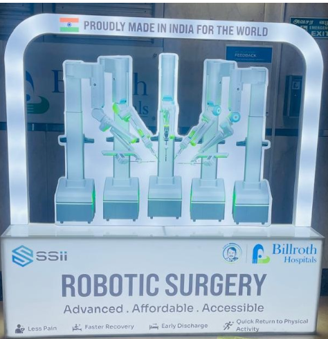
What Is Robotic Hernia Surgery?
Robotic hernia surgery is a minimally invasive procedure performed using a robotic surgical system that enhances a surgeon’s capabilities. While the robot does not perform the surgery on its own, it gives the surgeon extraordinary control through:
3D HD magnified vision (10–15x magnification)
Wristed instruments that rotate 540°
Ultra-precise movements with tremor filtration
This makes even complex hernia repairs smoother, safer, and more accurate.
Why Robotic Surgery for Hernia Repair?
Robotic platforms have transformed hernia surgery by overcoming limitations of traditional laparoscopy. Benefits include:
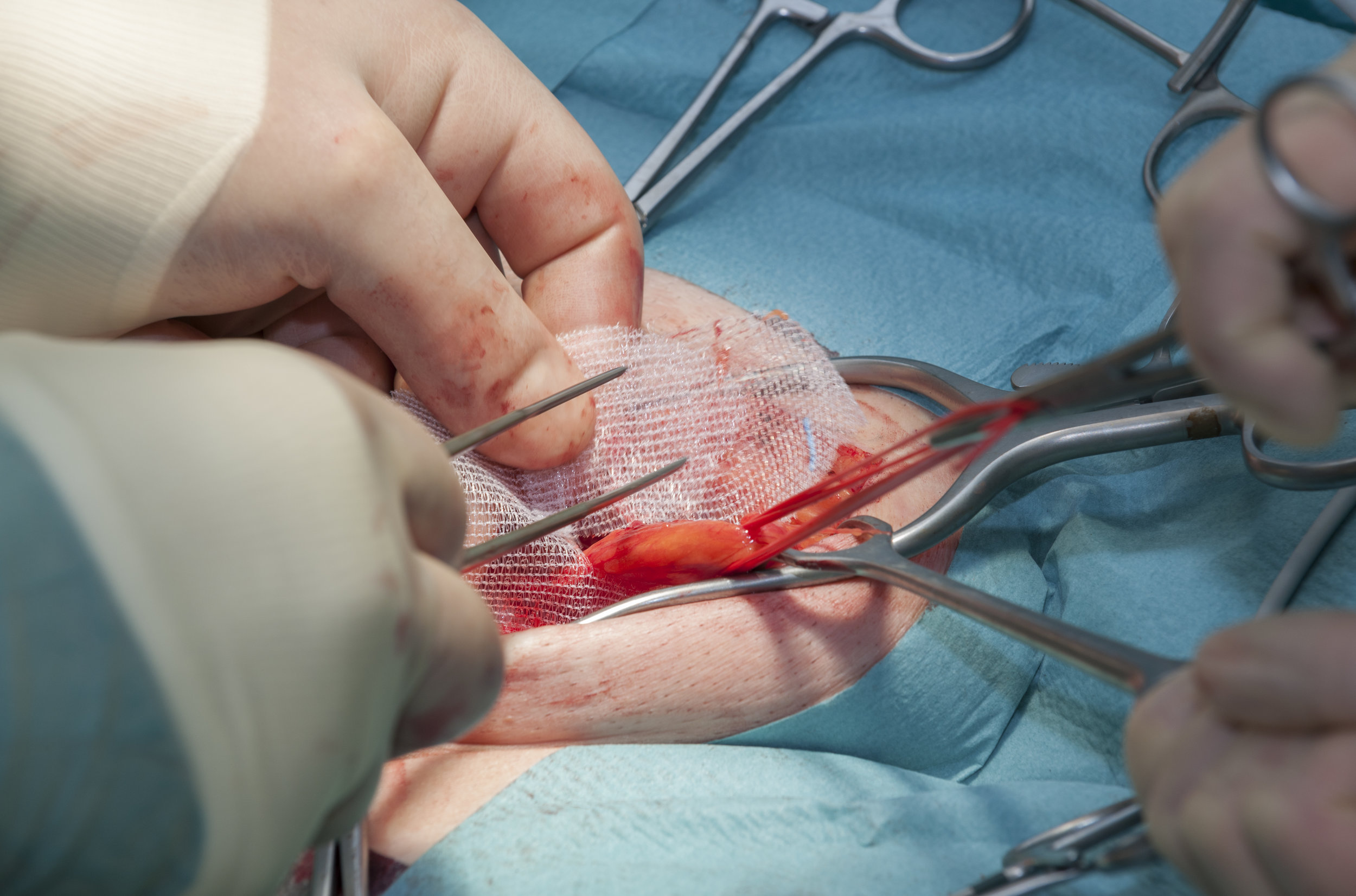
1. Greater Precision in Mesh Placement
Accurate mesh placement is crucial for long-term success. Robotic instruments allow meticulous dissection and suturing, reducing recurrence risks.
2.Less Pain and Faster Healing
Because the incisions are smaller and tissues are handled more gently, patients often experience:
Less post-operative pain
Reduced need for pain medication
Faster return to routine activities
3.Superior for Complex or Recurrent Hernias
Robotic surgery offers unmatched access and visualization, making it ideal for:
Large inguinal hernias
Ventral and umbilical hernias
Incisional hernias
Recurrent hernias after previous surgery
4.Reduced Complications
Improved precision means lower chances of:
Nerve injury
Mesh misplacement
Infection
Chronic groin pain
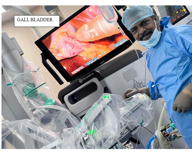
How Dr. Kumar Performs Robotic Hernia Surgery
Dr. Kumar follows a structured and safety-focused approach:
Step 1: Detailed Pre-operative Evaluation
Assessment of hernia size, location, previous surgeries, and patient’s health conditions.
Step 2: Minimally Invasive Access
Small holes are created to insert the robotic camera and instruments.
Step 3: Precision Repair
Using the robotic console, Dr. Kumar performs:
Gentle dissection of the hernia
Complete reduction of the hernia sac
Reinforcement with advanced mesh
Secure suturing for long-term support
Step 4: Quick Recovery
Most patients return home within 24 hours and resume normal work within 3–5 days.
Advantages for Patients
State-of-the-art robotic technology
Expertise in complex abdominal wall reconstruction
Standardised protocols for pain-free recovery
High success rates with minimal recurrence
Personalised care throughout the journey
Who Is an Ideal Candidate for Robotic Hernia Surgery?
Robotic repair is suitable for most patients, including those with:
✔ Inguinal (groin) hernias
✔ Umbilical hernias
✔ Ventral or incisional hernias
✔ Bilateral hernias
✔ Recurrent hernias
✔ Obesity or diabetes (safer than open surgery)
Dr. Kumar- Billroth Hospitals evaluates each patient individually to recommend the best approach.
Recovery After Robotic Hernia Surgery
Most patients experience:
Minimal pain
Quick mobility
Tiny scars
No restrictions on long-term activity
Return to office in a few days
Gym and workouts by 3–4 weeks (as advised)
Following Dr. Kumar’s enhanced recovery protocol ensures smoother healing.
Why Choose Dr. Kumar for Robotic Hernia Surgery?
Dr. Kumar is recognized for:
Expertise in advanced Laparoscopic and Robotic hernia repairs
High-volume experience with excellent outcomes
Focus on HERNIA Speciality
Impetus on Patient comfort and safety
Evidence-based techniques
Compassionate, personalised care
He offers one of the most comprehensive hernia care programs in Chennai.
Book a Consultation
To know if Robotic Hernia Surgery is the right choice for you, book an appointment with Dr. Kumar – Advanced Laparoscopic and Robotic Hernia & Abdominal Wall Surgeon, Chennai.